Appendix 1
Cellular physiology of absorption of
ORS solution
(Not recorded)
Figure 10 - Electrolyte, Glucose and Water
Transport in Small Intestine
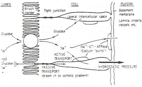
The reason that glucose greatly increases the absorption
of water and salt is because of a 'linked absorption
mechanism' in the enterocytes, the cells lining the small
bowel.
This means that in the presence of a substance like
glucose, sodium passes into the cells much more easily.
Figure 10 is a diagram of a single enterocyte in the
small bowel. In the centre is one complete enterocyte
and parts of two more. On the right are the
subendothelial layers of the lamina propria. The
absorption mechanism is in the brush border of the enterocyte; it takes up one molecule of glucose and one
ion of sodium and passes them into the cell together.
From there the glucose goes into the intercellular or
subcellular spaces and enters the circulation. The
sodium ions are transported by an active enzyme
mechanism which is sodium-potassium ATPase on the
base and lateral walls of the enterocytes. This "sodium
pump" as it is known, forces the ions into the lateral
intercellular spaces, where a high osmotic tension builds
up. This is one of the factors which then causes an
anatomical valve-like mechanism, the tight junction,
between enterocytes, to open up and take in larger
volumes of water, glucose and sodium from the lumen
of the bowel. Hydrostatic pressure then builds up in the
intercellular spaces and the fluid is forced into the
deeper layers and enters the circulation.